Undertake a full visual examination of the eye to be fitted, including measurements from the slit lamp, keratometer and corneal topography.
Select the optimum central base radius lens that gives a slightly steeper clearance than the corneal apex.
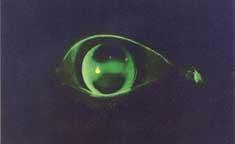
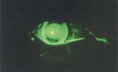
Should small air bubbles be observed anywhere under the lens, this would suggest the initial lens is too steep. A flatter fit should then be tried. If the lens fails to locate well or moves excessively, it may be found to be loose suggesting a steeper diagnostic lens should be tried.
Optimum Fit – NB Bridging
Good Fit – No Bubbles
ASSESSING THE PARACENTRAL AREA
To assist in fitting this area, the following questions and answers may be helpful, when the diagnostic lens is in situ.
Is there excessive stand-off in the paracentral area? Yes – order decreased lift. No – go to Question 2.
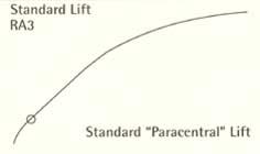
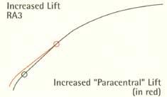
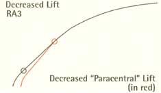
Is there excessive pressure in the paracentral area? Yes – order increased lift – see RA3 diagrams. No – go to Question 3.
Location – is the lens locating centrally? Yes – go to Question 4. No – Assess diameter – Increase diameter if riding low and decrease diameter if riding high.
Graft patients – Is there adequate applanation of the cornea (the bridging effect) with the diagnosis? Yes – move on to the RA zone. No – Re-assess the angle of the paracentral area. Either increase or decrease the lift to achieve the bridging effect – see diagram RA3.
Note: If required, altering the inclination of the tangential slope will assist in achieving good centration and applanation of the cornea.
To increase / flatten the paracentral area, order increased lift.
To decrease / steepen the paracentral area, order decreased lift.
Changes to the optic zone will also affect the paracentral area.
Increasing the optic zone will steepen the paracentral area.
Reducing the optic zone will flatten the paracentral area.
Important Note: Increased lift refers to the change to the paracentral area only.
REVERSE ASPHERIC GEOMETRY
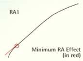
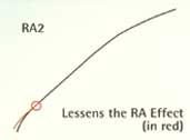
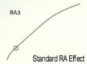
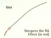
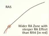
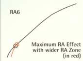
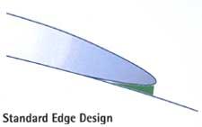
This third area requires assessment by fluoresein and slit lamp on the effect that the reverse aspheric periphery has on the peripheral profile. Refer to diagrams RA1 to RA6.
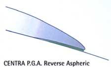
The standard Reversed Aspheric Geometry may be varied by the reference to the RA numbers. Of special interest, the PGA lens has a wider peripheral zone width, incorporated into its reverse aspheric design that is previously used in many contact lens’ designs.
The peripheral zone design creates a cushion of tears beneath the lens periphery for it to rest on. Centra PGA makes for greater overall patient comfort and reduces dust being trapped beneath the lens periphery.
FITTING
A diagnostic fitting set is essential for the successful application of this lens. The set consists of two systems:
For post grafts and high astigmatism: Base curves 7.0 – 8.6mm, diameter 10.1mm, optic zone 7.0.
For keratoconus and astigmatism: Base curves 5.3 – 7.6mm, diameter 9.0mm, optic zone 5.75
As noted, there is flexibility within the diameters and base range outside the standard parameters.
PER CASE PLAN
To make used of the advantages of the per case plan, CLC requires the use of a Centra trial set. These sets can be purchased or loaned. If the initial lens has been selected after use of a Centra fitting set and the final Ex lens is unsuccessful, CLC will issue a credit for 50% of the per lens price.
Price variances are defined as:
No exchange or refund for unsuccessful fits
with use of Centra Trial Set
+/- allows 2 exchanges per order
WARRANTY
RGP lenses come with a 30-day manufacturing warranty for defects. For an unsuccessful fit CLC will issue a 50% credit of the per lens price.